How We See
Eyeglasses and a visual test chart (DDurrich, iStockphoto)
Eyeglasses and a visual test chart (DDurrich, iStockphoto)
How does this align with my curriculum?
Learn about how human vision works as well as some common types of vision problems.
How We See
Vision
Rods & Cones
Cones
How the Brain Works With Our Eyes
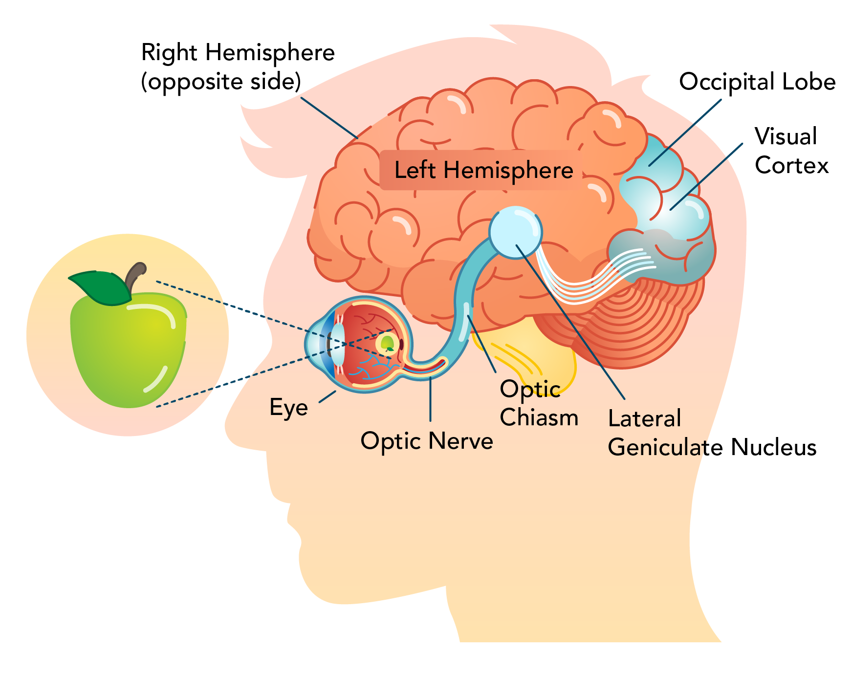
Image - Text Version
Shown is a colour illustration of a human head, showing how the eye and brain see a green apple.
The head is shown from the side, looking toward the left edge of the illustration. A green apple is left of the head, in front of the eye.
Two dotted, diagonal, straight lines lead from the top and bottom of the apple, into the eye. This is a pink sphere with clear, round, raised structures at the front, labelled "Eye."
The lines cross over each other in the middle of the pink sphere. A smaller, upside down image of the apple appears on the back inside wall of the eye. A tube leading from the back of the eye to the brain is labelled "Optic Nerve." A small, pale blue area halfway along the nerve is labelled "Optic Chiasm."
The brain is depicted as a pink, lumpy, oval structure. The visible side is labelled "Left Hemisphere." A line at the top edge leads to a label that reads, "Right Hemisphere (opposite side)."
The optic nerve leads to a pale blue circle near the centre of the brain. This is labelled "Lateral Geniculate Nucleus." From here, white curving lines fan out toward a region in the back upper part of the brain.
This area is coloured in pale blue. The top left part of it is labelled "Occipital Lobe." The lower right part is labelled "Visual Cortex."
Did you know?
The left side of the brain is called the left hemispheresand the right side of the brain is called the right hemisphere.
Vision Impairments
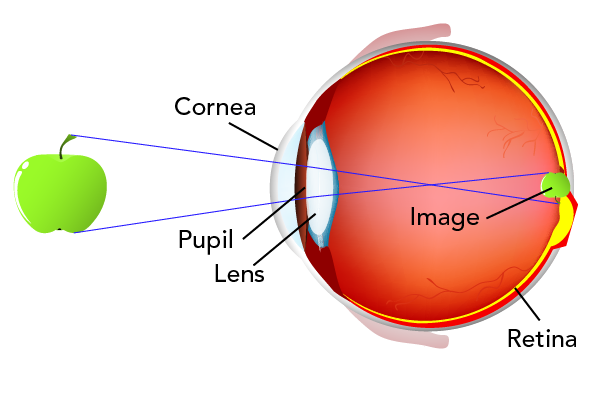
Image - Text Version
Shown is a colour illustration of an image of an apple formed in an eye with normal vision.
The eye is shown from the side, as if cut in half. It is shown as a pink sphere, with the cornea, pupil and lens labelled on the left. The front of the eye is facing left, where a green apple is in front of the eye.
Straight, blue, diagonal lines lead from the top and bottom of the apple, getting closer as they move towards the eye. These lines travel through the cornea, pupil and lens. They cross over each other in the centre of the pink sphere of the eye.
The crossed lines move apart again until they hit the retina at the back of the eye. Here, another, smaller green apple is shown upside down.
Near-sighted (Myopia)
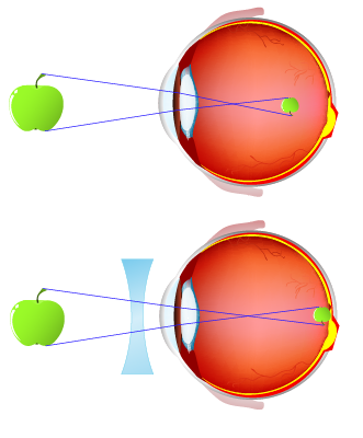
Image - Text Version
Shown are two colour illustrations of an image of an apple formed in an eye with myopia, and and the same eye through a biconcave lens.
In both illustrations, the eye is shown from the side, as if cut in half. It is shown as a pink sphere, with the cornea, pupil and lens labelled on the left. In front of the eye is a green apple.
Straight, blue, diagonal lines lead from the top and bottom of the apple, getting closer as they move towards the eye. These lines travel through the cornea, pupil and lens.
In the top illustration, the lines cross over each other near the front, inside the pink sphere of the eye.
The crossed lines move apart again until they meet an image of a smaller, upside down green apple. This image is not on the retina, but in the middle of the pink sphere, near the back.
In the bottom illustration is a blue lens with two edges that curve inwards. It is placed between the apple and the eye. Here, the diagonal blue lines leading from the apple cross over each other in the centre of the pink sphere. The image of the smaller, upside down apple is formed on the retina, on the back wall inside the eye.
Far-sighted (Hyperopia)
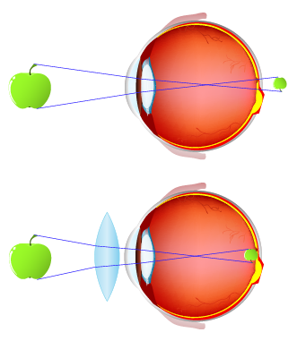
Image - Text Version
Shown are two colour illustrations of an image of an apple formed in an eye with hyperopia, and and in the same eye through a biconvex lens.
In both illustrations, the eye is shown from the side, as if cut in half. It is shown as a pink sphere, with the cornea, pupil and lens labelled on the left. In front of the eye is a green apple.
Straight, blue, diagonal lines lead from the top and bottom of the apple, getting closer as they move towards the eye. These lines travel through the cornea, pupil and lens.
In the top illustration, the lines cross over each other near the back, inside the pink sphere of the eye.
The crossed lines move apart again until they meet an image of a smaller, upside down green apple. This image is not on the retina, but beyond the back wall of the eye, on the white background of the illustration.
In the bottom illustration is a blue lens with two edges that curve outwards. It is placed between the apple and the eye. Here, the diagonal blue lines leading from the apple cross over each other in the centre of the pink sphere of the eye. The image of the smaller, upside down apple is formed on the retina, on the back wall inside the eye.
Presbyopia
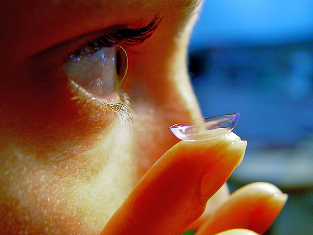
Contact Lenses
Image - Text Version
Shown is a person holding a clear, round lens on one fingertip, close to their right eye.
The lens is in the foreground of the photograph. It looks like a tiny clear glass cereal bowl. It is small enough to sit on the tip of the person's finger.
In the background, the right side of the person's face takes up more than half the image. Their eye is open and they look towards the right edge of the photograph. They are holding the lens up, just in front of their eye.