Ultrasonography
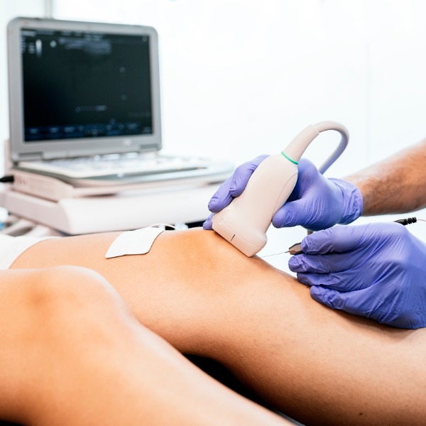
Ultrasound treatment on knee (santypan, iStockphoto)
Ultrasound treatment on knee (santypan, iStockphoto)
How does this align with my curriculum?
Learn about the history, function, uses, benefits and risks of Ultrasonography as a medical imaging technology.
Ultrasonography
Ultrasonography uses ultrasound to create images of soft tissues such as tendons, muscles, joints, blood vessels and internal organs. Unlike CT scans which use x-rays and MRI machines which use radio waves, ultrasound devices use sound waves to create the images.
History
Two researchers are particularly important in the history of medical ultrasound. Doctor Karl Theodore Dussik of Austria published the first paper on medical ultrasound in 1942. It was based on his research about the transmission of ultrasound through the brain. Professor Ian Donald of Scotland developed some of the first practical technology and applications for ultrasound in the 1950s.
How it Works
Ultrasound imaging devices take pictures using sound. Ultrasound are sound waves that have a very high frequency. The frequency of sound waves is measured in Hertz (Hz). Humans are able to hear sounds between a frequency of 20 to 20 000 Hz. Ultrasound uses frequencies between 2 000 000 and 17 000 000 Hz. Unlike sound waves that we normally hear which travel through air, ultrasound waves are mechanical waves that travel through a solid or liquid. This is why a gel is applied when a person gets an ultrasound. The gel helps the sound travel into the person.

One of the main parts of an ultrasound-imaging device is the transducer. The transducer is the instrument that the sonographer holds on the patient’s body. A sonographer is the technician doing the ultrasound imaging. A sound wave is produced by the transducer, which then travels through tissue at a constant speed of 1 540 m/s. Body tissues and structures will absorb, reflect or refract the sound wave.
Once the sound wave returns back to the transducer, it is converted from sound energy into electrical energy. The electrical energy is converted into pixels by the ultrasound machine and displayed as a video on a monitor. Soft tissue or organs show up on the monitor as shades of grey. Fluid and blood are black and bone is white. There are different types of transducers than can be used depending on the internal tissue or organ being imaged. A high frequency transducer is used to image surface structures with greater detail. A low frequency transducer is used to image structures deep within the body, but these images have less detail.
Did you know?
A pixel is the smallest element of an image.
Uses
Ultrasound is used to image many internal structures – even babies! Ultrasound imaging is widely used on pregnant women. This is called obstetric sonography.

Ultrasound is also used as a form of treatment. Ultrasound can help sprains and strains to heal faster. Focused high-energy ultrasound pulses are used to break up kidney stones and gallstones through a process known as lithotripsy. In addition to medical uses for humans, ultrasound has many other uses, such as in veterinary medicine for diagnosing and treating animals. They are used in motion-sensors, jewelry cleaners, humidifiers and many other devices. Who knew ultrasound technology was used for so many things!
Benefits & Risks
Ultrasound imaging is very safe to use and does not appear to cause any negative effects. It is also relatively inexpensive and quick to perform. Ultrasound scanners are portable and can be taken into places such as intensive care units. This avoids potential danger caused by moving critically ill patients to the radiology department where x-ray machines, CT scanners and MRI machines are located. The real-time moving images are also very useful in terms of diagnosing and treating illnesses.
Learn More
Ultrasound medical imaging | Mechanical waves and sound | Physics (2014)
This video by Khan Academy explains how and why ultrasound is used in medicine, and the information behind the images produced.
Ultrasound | GCSE Physics (2015)
This video by Doodle Science illustrates how ultrasound is used to investigate many materials, and the equation used to get results.
References
JennActon. (n. d.). The History of Ultrasound. TimeToast.com