Brain Architecture
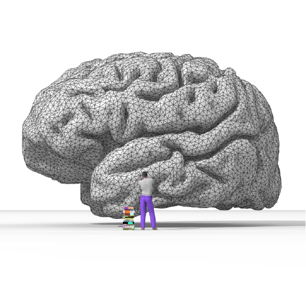
Rendering of the human brain (Nicolas Rougier, Wikimedia Commons)
Rendering of the human brain (Nicolas Rougier, Wikimedia Commons)
How does this align with my curriculum?
The human brain has many different parts. Each one plays a different function. But they all work together to manage complex thoughts, feelings and behaviours.
The human brain is one part of the nervous system. That’s the control system that sends instructions to all the other parts of your body.
Your brain has many different parts. Each one plays a different function. But they all work together to manage complex thoughts, feelings and behaviours.
Brain Cells
Like the other organs in your body, your brain is made up of cells. There are many different types of brain cells. Each one has a specific function.
Have you heard of neurons? They’re the most common type of brain cell. The average human brain contains about 86 billion neurons. And each one is connected to at least one other neuron forming close to 100 trillion connections in the brain.
At one end, each neuron has a cell body surrounded by dendrite branches. An axon connects the cell body to the axon terminal at the other end of the neuron. Schwann cells surround the axon. They provide a fatty coating called the myelin sheath.
Neurons transmit electrical signals called impulses. They flow through the axon from the dendrites and the cell body toward the axon terminal. Impulses jump from the axon end of one neuron to the dendrite end of the next. To do this, the signal has to cross a tiny gap called a synapse. Travelling from neuron to neuron, impulses move between brain regions at different speeds.

Regions of the Brain
On average, an adult human brain weighs about 1 300 grams. It uses about 20% of the body’s energy. The brain helps coordinate all of the body’s internal and external actions. Without your brain, you wouldn’t be able to sneeze, kick a ball or send a text.
Misconception Alert
People often say that humans only use 10% of their brain power. In fact, studies show that most of your brain is active at all times, including when you’re resting or sleeping!
The brain has three main parts. They’re the cerebrum, the cerebellum and the brainstem.
- The cerebellum helps fine-tune your muscle movement. For example, it helps control balance, posture and motor learning.
- The cerebrum is the largest part of the brain, spanning both the left and right hemispheres. It sits on top of the cerebellum and the brainstem. Many of your body’s higher functions rely on the cerebrum. For instance, it controls touch, vision, hearing, speech and fine motor skills. You also need your cerebrum to interpret emotions, solve problems and learn.
- The brainstem connects the base of the brain to the spinal cord. It helps coordinate the brain’s communication with the rest of the body. The brainstem also helps coordinate involuntary actions like breathing and heart rate.

The cerebrum has four main sections called lobes.
The frontal lobe is located at the front of the brain. It’s responsible for things like critical thinking, planning, motivation, feelings of reward and self-awareness. The frontal lobe also includes the motor strip. This brain region helps control body movement and contains the speech centre (Broca’s area).
The parietal lobe sits near the back of the brain. It’s located behind the frontal lobe and in front of the occipital lobe. The parietal lobe is responsible for sensing proprioception. In other words, it helps you understand the space around your body. It also contains the sensory strip. This brain region helps you sense things like pain and temperature.
The occipital lobe is found at the back of the brain. It contains the visual cortex, which allows you to interpret colour, light and movement. The two sides of the visual cortex process images contralaterally. That means what you see through your right eye is processed on the left side of your brain. And what you see through your left eye is processed on the right side of your brain.
The temporal lobe runs the full width of your brain, behind your temples. It’s responsible for learning, memory, understanding language (Wernicke’s area) and organization.

Inside the Brain
The brain contains both grey matter and white matter. Grey matter is located in the cerebral cortex. That’s the outer layer of the cerebrum. White matter is found closer to the centre. It’s made up of neurons with long axons that help connect different parts of the brain together.
The corpus callosum is a bridge of nerve fibres that connects the brain’s two hemispheres. It lets the hemisphere communicate with each other, so they can coordinate activity.
The thalamus is located deep inside the brain, above the brainstem. It coordinates all incoming sensory information. The thalamus also helps you feel pain, pay attention and remember things.
The hypothalamus sits just below the thalamus. It’s the control centre for the autonomic nervous system. This system controls what happens automatically in your body. For example, your breathing and the beating of your heart are controlled by the autonomic nervous system. The hypothalamus also links the nervous system to the endocrine system. This system is responsible for creating brain hormones and controlling the pituitary gland.
The pituitary gland is found below the hypothalamus. As part of the endocrine system, it plays a key role in human development. In fact, the pituitary gland is the control centre for all other hormone glands in the body.
The epithalamus is located behind the hypothalamus. The epithalamus contains the pineal gland which helps regulate the body’s internal clock by secreting a hormone called melatonin. This regulates your body’s circadian rhythms. They help you know when to wake up and when to go to sleep.
 corpus callosum, (2) thalamus, (3) hypothalamus, (4) pituitary gland, and (5) pineal gland")
Memory
Four parts of the brain are closely associated with memory. They are the amygdala, the hippocampus, the cerebellum and the prefrontal cortex.
The amygdala mainly regulates fear and aggression. It helps store memories related to these emotions. The amygdala recognizes these memories based on the higher levels of stress hormones produced when you feel afraid or angry. It also helps transfer new information into long-term memory.
The hippocampus is involved in recognition and spatial memory. This part of the brain also helps give memories meaning and connect them to other memories. Like the amygdala, the hippocampus helps transfer new information into long-term memory.
The cerebellum and the prefrontal cortex help consolidate procedural memory, motor learning and conditioned responses. Different parts of the prefrontal cortex are associated with storing and retrieving information.
 amygdala, (2) hippocampus, (3) cerebellum and (4) prefrontal cortex")
The Reward System
The reward system is a group of brain structures that get activated whenever you experience something rewarding.
Dopamine is a chemical neurotransmitter in the brain. It helps carry messages across the synapses that separate neurons. When the brain senses a rewarding stimulus, it releases more dopamine. This gives you a pleasant feeling. The brain pathway most often associated with reward is the mesolimbic dopamine pathway. It consists of three structures:
- Ventral Tegmental Area (VTA): This part of the brain receives information from the brainstem. Then it sends signals to the nucleus accumbens and the striatum.
- Nucleus Accumbens: This structure is responsible for feelings of pleasure, motivation and learning through reinforcement. It also plays a role in the coordination of movement.
- Striatum: The striatum regulates processes like action planning, motivation and decision-making.
The brain is a complex organ that plays an important role in shaping your behaviours, decisions and personality. Although each part may have a specific role, all of the parts must work together to help you form thoughts and feelings and carry out actions.
References
Editors of Encyclopaedia Britannica. (2018, December 3). Schwann cell.
Han, S. (2018, July 20). What are neurons?
Hines, T. (2018, April). Anatomy of the Brain. Mayfield Brain & Spine.
Lumen. (n.d.). Parts of the brain involved with memory.
Murrel, D. (2018, February 27). What percentage of our brain do we use?
Neuroscientifically Challenged. (n.d.). Mesolimbic pathway.
Spinal Cord. (n.d.). Occipital lobe.