Introduction to Bacteria
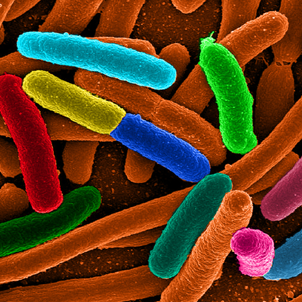
Assortment of the bacteria Escherichia coli (Mattosaurus [Public domain], Wikimedia Commons)
Assortment of the bacteria Escherichia coli (Mattosaurus [Public domain], Wikimedia Commons)
How does this align with my curriculum?
What exactly are bacteria? What types of bacteria are there? And are they really as bad as people think they are?
Bacteria have been around for a very long time. In fact, they are the oldest known forms of life on Earth! The earliest fossils we know are of prokaryotes. That’s a group of organisms that includes bacteria. These fossils are about 3.5 billion years old!
Since then, bacteria have evolved into a wide variety of types. They have also adapted to many different environments. They can live inside the human body, at the North Pole, and even at the bottom of the ocean!
Did you know?
There are more bacteria living in your mouth than there are people who have ever lived on Earth!
Size of Bacteria
Bacteria are single-celled organisms. This means that each bacterium is made up of only one cell. This is very different from humans. Our bodies are made up of trillions of cells.
Bacteria are much smaller than human cells. Bacterial cells are between about 1 and 10 μm long. And most of them are only 1 to 2 μm in diameter. 1 μm, or micrometre, is 1 000 times smaller than a millimetre. That is much smaller than the human red blood cell, which is about 7 μm in diameter.

Image - Text Version
Shown is a colour illustration of cell and cell parts along a scale from one nanometre to one centimetre, labelled with the equipment needed to view them. The scale starts on the left at one nanometre. Above this marking is an illustration labelled "Small Molecule". This consists of multicoloured spheres connected by white sticks. Between ten and 100 nanometres is an illustration labelled "Virus." This looks like a clear teal gem sitting on a purple pole with thin spider legs around the base. Over the one micrometre mark is an illustration labelled "Bacterium." This is a green capsule shape that looks a bit like a pickle. Over the ten micrometres mark is an illustration labelled "Animal Cell." The is flat, round, and blue with purple shapes inside. Over the 100 micrometres mark is an illustration labelled "Plant Cell." This is a soft, rounded green rectangle with pointed corners and yellow and dark green shapes inside. It looks a bit like a pillow. Below the scale are two thick, coloured stripes. The top one is yellow and labelled "Electron Microscope." This goes from smaller than one nanometer to the 100 micrometres mark. The lower stripe is blue and labelled "Light Microscope." This runs from halfway between the 100 nanometre and one micrometre marks to halfway between the one millimetre and one centimetre marks.
Even though they are small, bacterial cells have many different parts.
Bacterial Structure
Click on the numbers to learn more about the parts of a bacterial cell
Click here to access screen-reader friendly PDF
Classification of Bacteria
There are millions of different types of bacteria in the world. That’s why it’s important to have a way to classify them. Scientists usually classify bacteria using two characteristics:
- The thickness of its cell wall
- Its shape
Scientists use Gram staining to measure the thickness of cell walls. To do this, they stain bacteria with a dye called crystal violet. Thick cell walls keep the violet colour of the dye. Thin cell walls do not.
- Gram Positive bacteria have thick cell walls. They appear blue or purple when dyed.
- Gram Negative bacteria have thin cell walls. They appear pink or red when dyed.
Bacteria Shapes
Bacteria are classified into three major groups based on their shape. The first group is spherical (cocci), the second is rod-shaped (bacilli), the third is spiral-shaped and others.

Image - Text Version
Shown is a colour illustration of ten different bacteria, grouped into three columns. The title, "Shapes of Bacteria" is in white block letters on a red rectangle across the top. Starting on the left, the first column is titled "Spherical (cocci)" in green. The cells below are also green. The first cell is two spheres stuck together in a row. This is labelled "Streptococcus pnuemoniae: Causes pneumonia." The second cell resembles a worm. It is about 12 spheres squished together into a curved line. This is labelled "Streptococcus pyogenes: causes Strep Throat." The third cell is four spheres stuck together in a square shape. This is labelled "Micrococcus luteus: causes armpits to stink." The fourth cell in the group is spheres stuck together into a flat shape with five straight sides and one curved one. This is labelled "Staphylococcus aureus: can cause sinus infections and food poisoning." The middle column is titled "Rod-shaped (bacilli)" in orange letters. The cells below are illustrated in orange. The first cell in this column is four capsule shapes stuck together, end-to-end. This is labelled "Bacillus anthracis: causes Anthrax." The second cell is a single capsule shape with six long, squiggly legs. This is labelled "Salmonella enterica: causes Typhoid." The third cell in this group is a cylindrical shape with sharply angled yellow ends. This is labelled "Clostridium botulinum: causes Botulism." The right-hand column is titled "Spiral-shaped (and others)" in purple letters. The cells below are also purple. The first cell looks like a short, flexible tube with rounded ends and a long, squiggly tail. This is labelled "Vibrio cholerae: causes Cholera." The second cell is a slightly longer tube with four squiggly tails. This is labelled "Helicobacter pylori: can cause stomach ulcers." The third cell in the group is shown as two long, thin, wavy tubes. These are labelled "Treponema pallidum: causes Syphilis."
Are Bacteria Bad?
We often think of bacteria as being ‘bad’. That’s because some bacteria are pathogenic. This means they can make us sick. Lots of diseases and conditions are caused by bacteria. These include:
- tetanus
- typhoid fever
- tuberculosis
- strep throat
- anthrax
- food poisoning

Image - Text Version
Shown are three black and white photographs of bacterial cells, arranged in a row. The left photograph shows three large, uneven spheres and two smaller ones. Both have light grey, bubbly textured surfaces. They are scattered on a dark grey surface dotted with black circles. The centre photograph shows bright white tube or capsule shapes with one tail each. These float in dark grey space. The third photograph shows what looks like a thin, dark grey wire twisted into a vertical corkscrew shape. Several other, similar shapes are out of focus in the background. They stretch up from, or lie down on, a surface that looks like grey mud.
Did you know?
Researchers are starting to use bacteria to dye clothing. This could be a lot safer and more sustainable than traditional dyes.
But only a small fraction of the bacteria in the world cause us harm. In fact, many bacteria are helpful! For example, probiotic bacteria in our digestive system helps protect us from other bacteria.
Bacteria are all around you - even in your kitchen! Lots of food and beverages are made from bacteria. These include:

Image - Text Version
Shown are six colour photographs of foods, arranged in a grid. Starting on the top left, the first is a blue bowl full of thick white yogurt, with blueberries on top. To the right, the second is an oval-shaped, crusty loaf of bread with a paper label. The third photograph is filled with liquid chocolate. A spoonful of thick, glossy rich brown chocolate is held above the surface. On the bottom row, the first photograph shows a table piled with cheeses in many different shapes, colours and textures. The second is a white bowl full of soft, chopped cabbage leaves in a cream-coloured sauce, sprinkled with green herbs. The last photograph is a wide dish of large cabbage leaves in a red sauce.
Bacteria are diverse and interesting living things. People may think of them as things that make us sick, but they do a lot of good for us as well!
Learn More
The science of cheese (2020)
This page from Science Learning Hub explains the ancient biotechnology of cheesemaking and includes an animated video of how the molecular structure of milk changes during the process.
Structure of Bacteria | Cells | Biology | FuseSchool (2016)
This video (2:57 min.) from FuseSchool describes the structure of bacteria and why they are so important.
GRAM POSITIVE VS GRAM NEGATIVE BACTERIA (2019)
This video (3:09 min.) from Neural Academy explains the difference between gram positive and gram negative bacteria.
Bacterial Structure and Functions (2021)
This video (6:58 min.) from Osmosis.org goes into more depth about bacterial structure and function.
Bacterial Disease | Health | Biology | FuseSchool (2019)
This video (3:48 min.) from FuseSchool explores several types of bacteria that cause human illnesses.
A brief history of TB (2021)
This video (2:51 min.) from the Microbiology Society looks at the history of tuberculosis (TB), one of the world’s deadliest diseases.
References
Arizona State University. (2014, July 3). Microbes: The good, the bad, the ugly.
Bailey, R. (2019, August 20). Bacteria shapes. ThoughtCo.
Bruckner, M. Z. (n.d.). Gram staining. Carlton College.
Davidson, M. W. (2015, November 13). Bacteria cell structure. Florida State University.
Humm Kombucha. (2017). The difference between good bacteria and bad bacteria.
Microbiology Online. (n.d.). Bacteria. Microbiology Society.
Microscope Master. (n.d.). Unicellular organisms.