Radiography & X-ray Imaging
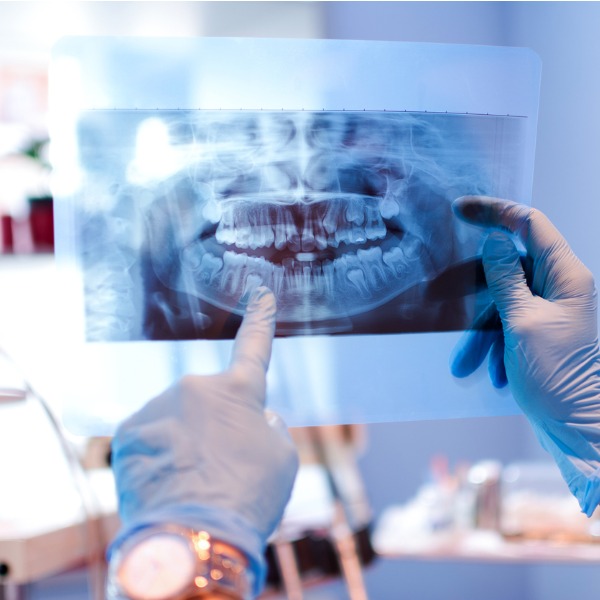
dental x-ray (Bojan89, iStockphoto)
dental x-ray (Bojan89, iStockphoto)
How does this align with my curriculum?
Learn about radiography and x-rays as a form of medical imaging technology.
There are many forms of imaging technology. Imaging technology involves using various materials and types of technologies to create images. An image (from the Latin word imago, meaning ‘a picture,’ or ‘likeness’) is a visual representation of a person, place or thing. It can be two-dimensional, such as a photograph, or three-dimensional, such as a statue. Some images are very short-lived, such as the image of your face in a mirror or the images on a TV screen. Other images are more permanent. Permanent images are known as fixed images or hard copies. Telescopes, cameras, microscopes and other optical devices are all used to capture images.
Radiography
Radiography is the creation of images using x-ray energy. Images created using this technique are called radiographic images (or radiographs). X-ray images are still pictures similar to photographs. Fluoroscopic images are moving pictures similar to movies. Tomographic images are visualized ‘slices’ of the body.
Medical Radiation Technologists (MRTs) are the professionals who create radiographic images. Several types of radiographic imaging are used in health care. MRTs need a good understanding of both biological sciences and physical sciences. The biological sciences they study are primarily human anatomy and physiology, including osteology. Osteology is the study of the bones that make up the human skeleton. They also study physical sciences, such as physics and chemistry. Through their studies they learn how x-rays are produced. They learn how the x-ray beam changes when it interacts with the tissue of a body to create an image. They also learn how the image is captured and how the image is stored.
X-ray Imaging
Imagine you were playing your favourite sport and right when you were about to be the star of the day, a kid from the other team crashes into you! After rushing to the hospital, where you get an x-ray of your bone, you might ask yourself, “What is an x-ray?”
History
X-rays were discovered on November 8th, 1895 by German physicist Wilhelm Conrad Roentgen while he was experimenting with cathode ray tubes. A cathode ray tube is a sealed tube with all the air removed. At one end of the tube is an electron gun and at the opposite end of the tube is a screen on which images can be viewed. Roentgen noticed that photographic plates made using a chemical called barium platinocyanide would fluoresce (glow) even when the cathode rays were blocked. What he discovered was a new type of electromagnetic radiation which he called x-radiation. He used the letter “x” because in mathematics “x” represents an unknown quantity. The term was later shortened to x-rays. The x-rays that Roentgen discovered are a highly penetrating form of electromagnetic radiation. He found that the rays could pass through soft human tissue, but could not pass easily through hard tissue, such as bone, or through metal, such as his wife’s ring.

He also found that the rays could make images on photographic film. For his pioneering work, Roentgen received many honours, including the first Nobel Prize for Physics in 1901. The unit that was used for a long time for measuring exposure to x-rays, R (roentgen), was named in his honour. Exposure to x-rays is now measured in Coulombs per kilogram (C/kg).
How it Works
An x-ray machine is essentially a specialized type of camera. Instead of having visible light expose the film and create an image, x-rays expose the film and create an image. Bone tissue, which contains a lot of calcium, absorbs a lot of x-rays, whereas soft tissues such as fat and skin do not. In an X-ray image, areas that contain less dense tissue appear darker while areas that contain denser tissue appear lighter. This is why bones and metal objects look white in x-rays.

This is the opposite of what we saw in Roentgen’s first x-ray image. That image was a positive image (image transferred from photographic film to paper) whereas the image above is a negative image (image on photographic film).
Uses
X-rays are not just for checking for broken bones. Your dentist uses x-ray imaging to check for cavities in your teeth. Mammographers use low-energy x-rays to do mammograms, which help doctors check the health of breast tissue. Airport security uses x-ray machines to check luggage for dangerous objects.

X-rays are also used as a form of non-destructive testing (way of testing for weaknesses without destroying the object). X-ray images can show hidden flaws such as tiny cracks or flawed welding in items such as pipelines.
Benefits & Risks
The benefit of x-ray imaging is that it is a very useful tool for looking at bones, as well as other hard tissues. This includes gallstones, which are crystals sometimes formed in the gallbladder. It also includes kidney stones, which are crystals sometimes formed in the kidneys. Other hard objects include cancerous tumours and accidentally swallowed objects, such as keys, coins and safety pins. On the negative side, x-rays are a form of ionizing radiation. This means that they can increase the risk of developing cancer. However, the amount of radiation exposure we get from dental x-rays or x-rays of broken bones is very small.
Learn More
How X-rays see through your skin - Ge Wang (2015)
This video by TED-Ed explores the discovery of x-rays, how they work, and how they are used in computed tomography.
X-Rays | Science Mission Directorate (2016)
This article with video (2:50) by NASA explains the characteristics of x-rays, where they’re found in the universe, how they’re used to investigate the sun, Martian rock, supernovas and the earth’s aurora.
References
Harris, T. (n. d.) How X-rays Work. HowStuffWorks.