Specialized Cells of the Leaf System
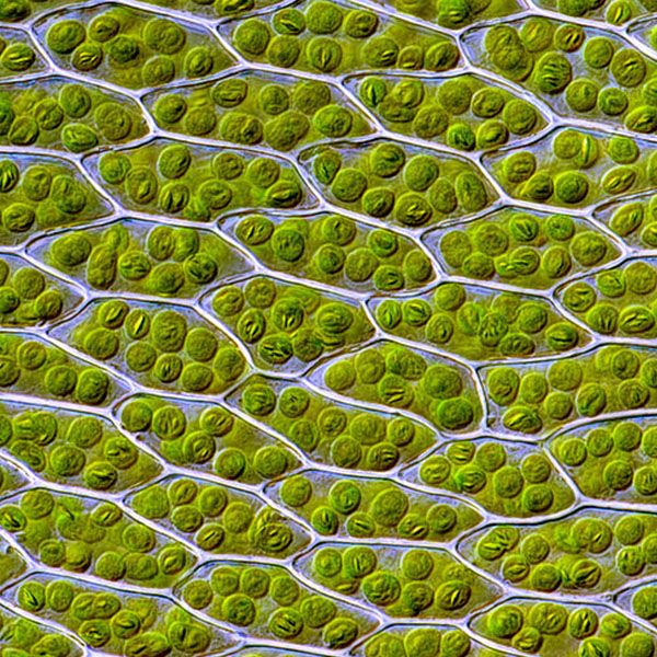
Plant Cells with Visible Chloroplasts

Plant Cells with Visible Chloroplasts (Des_Callaghan, Wikimedia Commons)

Plant Cells with Visible Chloroplasts

Plant Cells with Visible Chloroplasts (Des_Callaghan, Wikimedia Commons)

6.35

How does this align with my curriculum?

Share on:

Learn about the structure and function of the cells in leaves.

Leaves are essential to life on earth.

They can be tiny, like the leaves of the common water fern (Azolla filiculoides), which are just one millimetre in length. And they can be large, like the leaves of the raffia palm (Raphia regalis) that can grow to be 25 metres in length.

No matter the size, most green leaves take part in photosynthesis. This is the process in which plants convert light energy into sugars and oxygen. Photosynthesis allows plants to grow, providing food and oxygen for the whole planet.

Leaf Structure

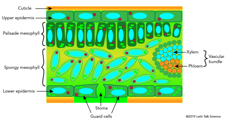
Leaves are complex organs made of several layers of tissue. These are the epidermis, the palisade mesophyll layer, the spongy mesophyll layer, and vascular bundles.

Cross-section of a dicot leaf showing its various tissues and anatomy
Cross-section of a dicot leaf showing its various tissues and anatomy (©2019 Let’s Talk Science).
Image - Text Version

Shown is a colour illustration of a cross-section of a leaf, showing the parts inside.

The top layer of the leaf is coloured in orange. This is labelled Cuticle with a black arrow.

The second layer is made up of horizontal, rectangular green shapes with rounded corners, in a straight row. Inside each rectangle is a bright greenish blue oval and a small red circle. The small red circle is always close to the narrow end of the oval, sometimes on the left, and sometimes on the right. This layer is labelled upper epidermis.

The third layer is made up of vertical, dark green rectangles with rounded corners. These vary in length, and they stand side by side in an uneven row. Inside the rectangles are bright greenish blue ovals and red circles, as before. There are also between one and four tiny, bright green rectangles in each dark green one, depending on its size. This layer is labelled Palisade mesophyll.

The background of the fourth layer is greenish yellow at the top, shaded to pale green at the bottom.

On the left of this area, lots of long pale green ovals float loosely in the space. Each oval has a smaller, bright greenish blue oval, and a tiny red circle inside it. This layer is labelled Spongy mesophyll.

On the right edge of the illustration, next to the Spongy mesophyll, is a large half-circle of small green circles, like a beaded necklace. Inside, the top part of the circle is filled with similar circles, in bright greenish blue. The bottom part is filled with round orange circles. The greenish blue circles are labelled Xylem and the orange ones are labelled Phloem. The words Xylem and Phloem together are labelled Vascular bundle.

The fifth layer is made up of a row of horizontal green rectangles, similar to the second layer. Each rectangle has a bright greenish blue oval and a red circle inside, but they also have three to five tiny, bright green rectangles each. This layer is labelled Lower epidermis.

  •  Theepidermis is the “skin” of a leaf. Sometimes there is also a cuticle outside the epidermis. This is a waxy layer that helps prevent water loss. This is especially important in dry regions. Leaves have an upper epidermis and a lower epidermis. The lower epidermis is located on the underside of leaves. This layer often has stomata  (singular is stoma). Most dicot plants have their stomata on the lower epidermis. This stops the plant from losing too much water through evaporation. This is because the lower epidermis is usually in the shade. Monocot plants like corn and grass can have stomata on both the top and bottom sides of leaves. This is because their leaves grow straight up, not parallel to the ground. This means evaporation happens on both the upper and lower epidermis.
  • The palisade mesophyll layer is just below the upper epidermis. It is made up of closely-packed, elongated cells. These cells contain chloroplasts. They perform most of the photosynthesis.
  • Vascular bundles are made up of xylem and phloem  cells. These cells carry water and nutrients throughout the plant. You can see them as the veins in leaves. The xylem is responsible for transporting water and nutrients from the soil to the leaves. The phloem transports sugars from the leaves to all other cells of the plant.
  • The spongy mesophyll layer is just below the palisade mesophyll layer. Its cells are irregularly-shaped and loosely packed. Cells in this layer usually contain few chloroplasts, especially in dicot plants. In this layer, air spaces between the cells are connected. They are also connected to the outside through the stomata. These are small pores, or holes on leaves.

Chloroplasts and Photosynthesis

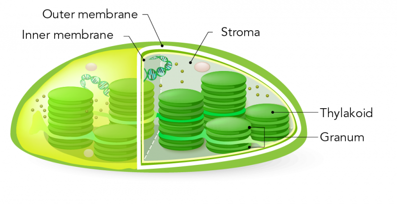
Chloroplasts are specialized organelles  that are only found in plant cells. Chloroplasts contain thylakoids. Thylakoids are the specific structures responsible for photosynthesis. Some thylakoids float freely in the fluid that fills the chloroplast. This fluid is called stroma. But most thylakoids are in stacks called grana. A single grana is called a granum. Grana resembles a stack of coins or pancakes.

Structure of a chloroplast
Structure of a chloroplast (Source: Let’s Talk Science using an image by ttsz via iStockphoto)
Image - Text Version

Shown is a colour illustration of a chloroplast, with a section of the outer surface cut away to show the structures inside.

The chloroplast is an oval shape, flat on the bottom and domed on top. A dark green layer around the outer surface is labelled Outer membrane. This layer is slightly wavy, so that it appears flexible. Inside that, a thinner, pale yellow layer is labelled inner membrane. 

The area of the membranes that is cut away shows the chlorophyll is filled with a clear, pale grey substance. This is labelled Stroma. The largest structures in the chlorophyll are seven stacks of dark green discs. A single disc is labelled Thylakoid. The stacks vary in height. One almost reaches the inner membrane at the top, with 9-10 thylakoids in the pile. The smallest stack has 4-5 thylakoids. A stack is labelled Granum.

Thylakoids have chlorophyll and other proteins in their membranes. Chlorophyll is a molecule that traps energy from the Sun. Plants use this energy to build sugar molecules, which the plant can later use as a source of energy.

Did you know?

Chlorophyll molecules reflect green light. This is why so many parts of plants appear green to us. Chlorophyll is one of several pigments that give plants their colour.

Stomata and Gas Exchange 

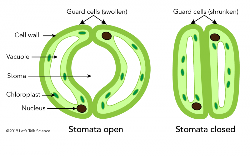
The stomata in a leaf’s surface are surrounded by specialized cells called guard cells. Guard cells regulate the opening and closing of stomata. They open by taking in water. This causes them to swell and take on a crescent shape. They close by releasing water. This causes them to shrink and take on a long, thin shape.

Stomata allow oxygen (O2) and carbon dioxide (CO2) to enter or leave the plant. This process is called gas exchange. The stomata also allow water vapour to leave the plant. This is part of the process of transpiration.

Structure of a chloroplast
Diagrams of open (left) and closed (right) stoma (©2022 Let’s Talk Science).
Image - Text Version

Shown is a colour illustration of open and closed stomata.

The left illustration is labelled Stomata open. It shows two long green cells curved into half circles, touching at the top and bottom, with an open space in the middle. The cells are labelled Guard cells (swollen), and the space is labelled Stoma.

The thick, dark green layer on the outside of the cells is labelled Cell wall. Inside each cell is a pale green layer, with tiny, dark green ovals in it. These ovals are labelled Chloroplast. A larger black oval sits in the bottom end of the left cell and the top end of the right cell. These are labelled Nucleus. At the centre of each cell is a large white area labelled Vacuole.

The right illustration is labelled Stomata closed. The two cells here are labelled Guard cells (shrunken). They look similar to the ones on the left, but the vacuoles are smaller, and the cells meet tightly in the middle, leaving no space (stoma).

How is the opening and closing of stomata regulated?

Each guard cell contains a large vacuole, a nucleus and chloroplasts. Generally, when the plant senses good conditions, like bright light or high humidity, its stomata open. This happens when guard cells swell with water through the process of osmosis. The reverse happens when stomata close in poor conditions.

Leaf Structure and Function (2021)
This video (5:52 min.) by BOGOBiology walks through the structure and function of each component, and connects each back to the process of photosynthesis.

The Chloroplast (2016)
This video (5:36 min.) by Boseman Science explains how the chloroplast in plants harnesses power from the Sun to form high energy molecules like glucose.

Structure Of The Leaf | Plant | Biology (2013)
This video (3:42 min.) by The FuseSchool, explains how plants use their leaves to make food through photosynthesis.

Leaf Stomata Lab (2018)
This video (6:24 min.) by Julian Buss shows how you can see the impression of stomata under a microscope using clear tape and nail polish.

References

Biology Pages. (n.d.). Gas exchange in plants.

Biology Pages. (n.d.). The leaf.

Enchanted Learning. (n.d.). Leaves and leaf structure.

Hyperphysics. (n.d.). Chloroplasts. Georgia State University.

Science & Plants for Schools. (n.d.). Measuring stomatal density.

SparkNotes. (n.d.). Plant structures.